About Equipment
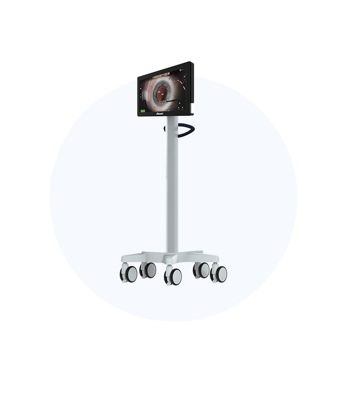
DMM
DIGITAL MARKER M
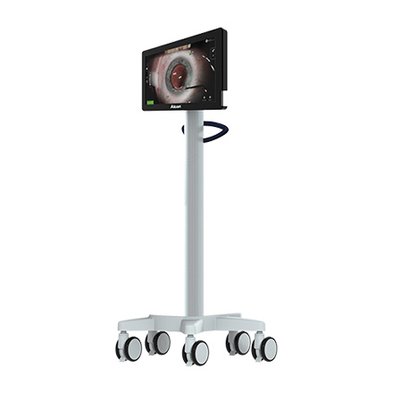
DMM is a device that provides guidance in the operating room using images taken by ARGOS and planning data.
01DMM offers guidance directly usable in the operating room based on the measurements and images obtained from the patient's eye captured by ARGOS. It ensures accurate guidance throughout the surgery, from the incision to lens emulsification and alignment of the intraocular lens, not only for calculations of the artificial lens and astigmatism done by ARGOS.
02Therefore, even if the patient moves their eye during surgery, it automatically tracks the position, reducing variables of refractive errors, and corrects the procedure to ensure the surgeon achieves the intended refractive outcome.
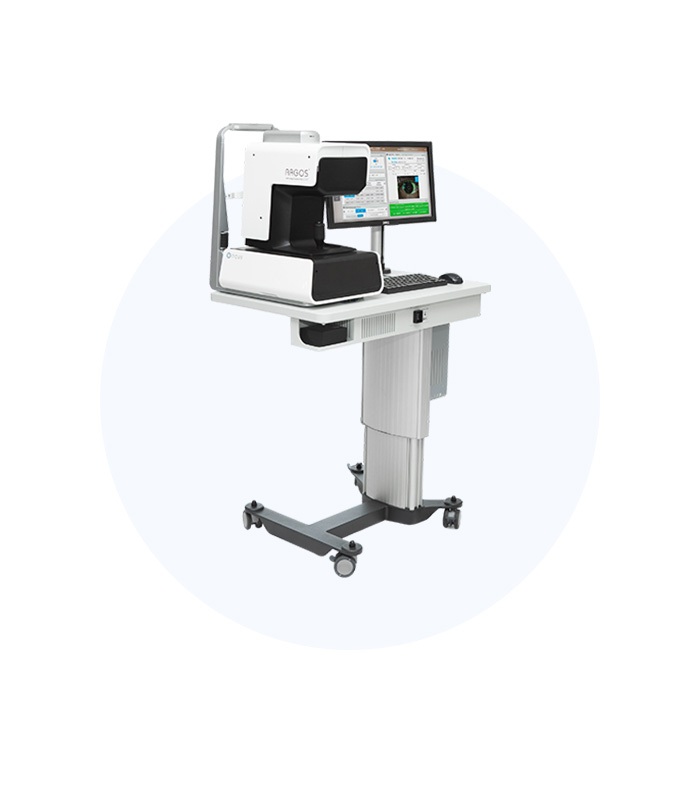
ARGOS
ARGOS BIOMETER
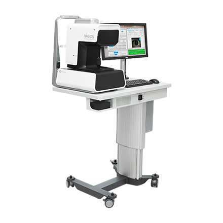
Argos is a corneal curvature radius measurement and biometric testing device that visualizes and measures the eye structure and calculates the necessary artificial lens.
01Utilizing the SS-OCT method, ARGOS measures the values needed for cataract surgery or refractive correction and performs calculations to determine the type and power of the artificial lens. Particularly, the segmented axial length measurement method used in the ARGOS device employs appropriate wavelength lasers for each eye measurement, resulting in more accurate test results.
02The newly integrated Image Guidance System in Alcon's ARGOS captures images of the eye before and after surgery, serving as a navigation tool alongside DMM during pre-operative planning and the surgery itself, thus helping achieve more accurate surgical outcomes for patients.
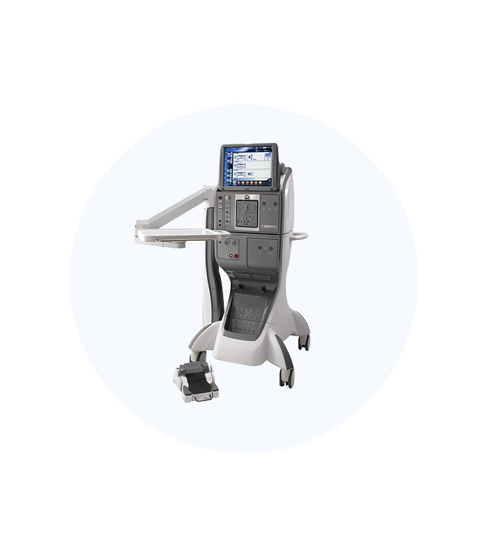
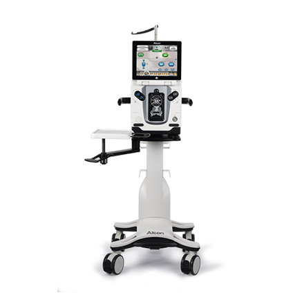
CONSTELLATION
CONSTELLATION
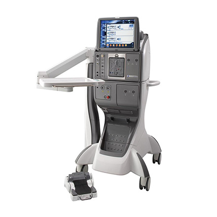
This surgical equipment is primarily used in vitrectomy, which constitutes a significant portion of retinal surgeries.
As an innovative device for retinal and cataract surgery, it allows for effective, safe surgeries with stable intraocular pressure and rapid vitrectomy speed.
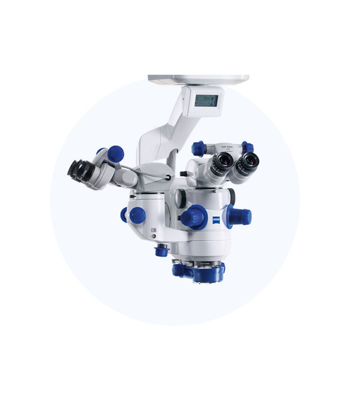
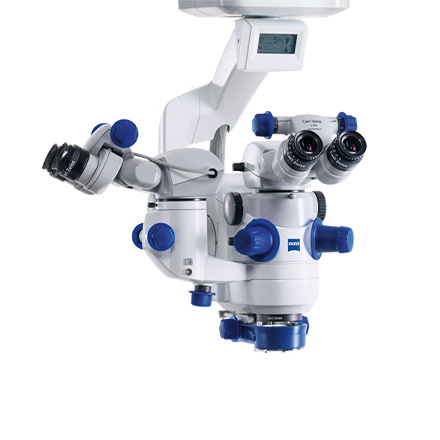
Lumera 700
Lumera 700
Previous microscopes lacked clarity, making it difficult to observe the interior of the patient's eye accurately. However, Lumera offers significantly higher clarity, enabling easier surgical procedures.
Additionally, while previous models illuminated only the cornea, Lumera uses three light sources to simultaneously illuminate the cornea, lens, and retina, allowing for three-dimensional observation.
Most importantly, even in cases of corneal opacity or severe cataracts, the high clarity allows for the successful execution of complex surgeries.
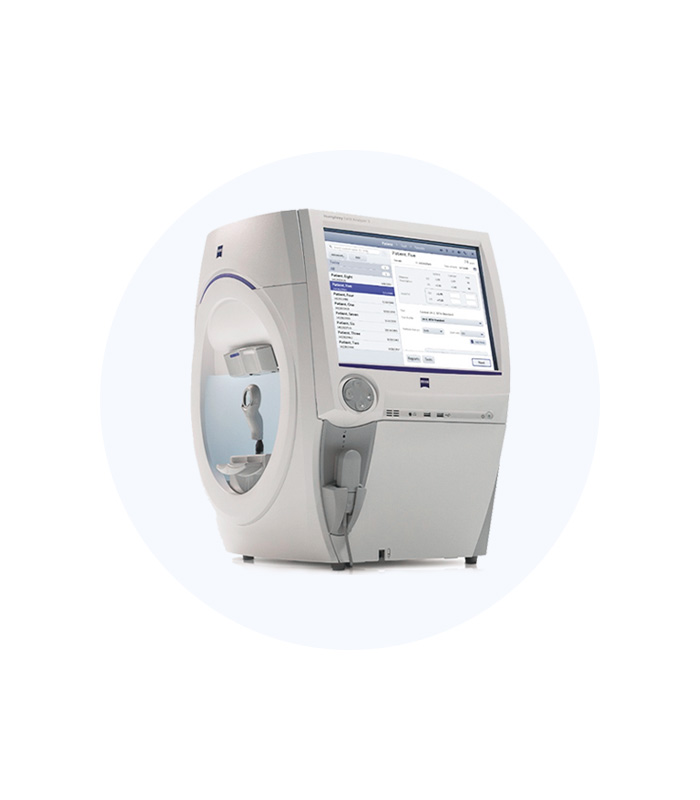
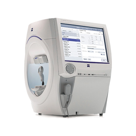
SIGNATURE PRO
SIGNATURE PRO
The visual field tester is the most accurate device for diagnosing glaucoma and retinal conditions, offering precise diagnostics and analysis of glaucoma progression.
01. SITA Method and Automatic AnalysisThis device adopts the SITA method, significantly reducing visual field test time. Furthermore, with Visual Field Index automatic analysis, it selectively analyzes glaucoma-related visual field damage.
02. GPA TesterEquipped with a GPA tester that automatically indicates glaucoma progression, it enables convenient and precise diagnostics.
03. Dynamic Automated Visual Field TestingDynamic automated visual field testing allows for rapid examination of non-glaucomatous conditions such as disability diagnoses or neurological diseases.
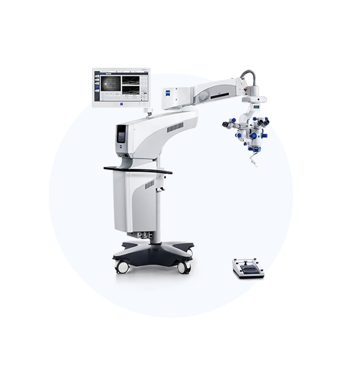
ZEISS CALLISTO EYE
ZEISS CALLISTO EYE
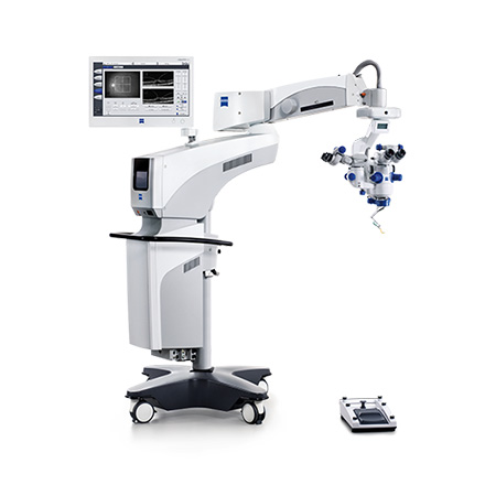
The ZEISS Cataract Suite Markerless consists of four products released by ZEISS: IOL Master, Forum, Callisto Eye, and Lumera Microscope, enabling faster and more accurate surgeries during toric and premium cataract procedures through a computer navigation system.
01. ForumThrough Forum, test results from various devices can be reviewed and quickly transmitted, and during surgery, it employs vessel matching with reference images taken from the IOL Master in real-time.
To achieve accurate vessel matching, obtaining a good reference image from the IOL Master is crucial, requiring the examination to capture as many vessels as possible. Given the characteristics of Asian patients, often having smaller or drooping eyelids, this requires the examiner's experience and time.
The ZEISS Cataract Suite Markerless allows for surgeries without the cumbersome procedures of placing sharp instruments on the patient’s eye pre-operatively or marking the eye during surgery, thereby providing faster and safer surgery for patients.
Through the computer navigation system, Callisto Eye can confirm where and how much to incise during Limbal Relaxing Incision (LRI) and guide the size and position of Continuous Curvilinear Capsulorhexis (CCC).
Additionally, during toric surgeries, it provides precise guidance on the axis where the astigmatism-correcting intraocular lens will be placed, minimizing axis errors to offer more accurate surgery for patients.
Callisto Eye's functions can be checked not only on a touch screen monitor but also in real-time through the eyepiece of the Lumera Microscope during the procedure.
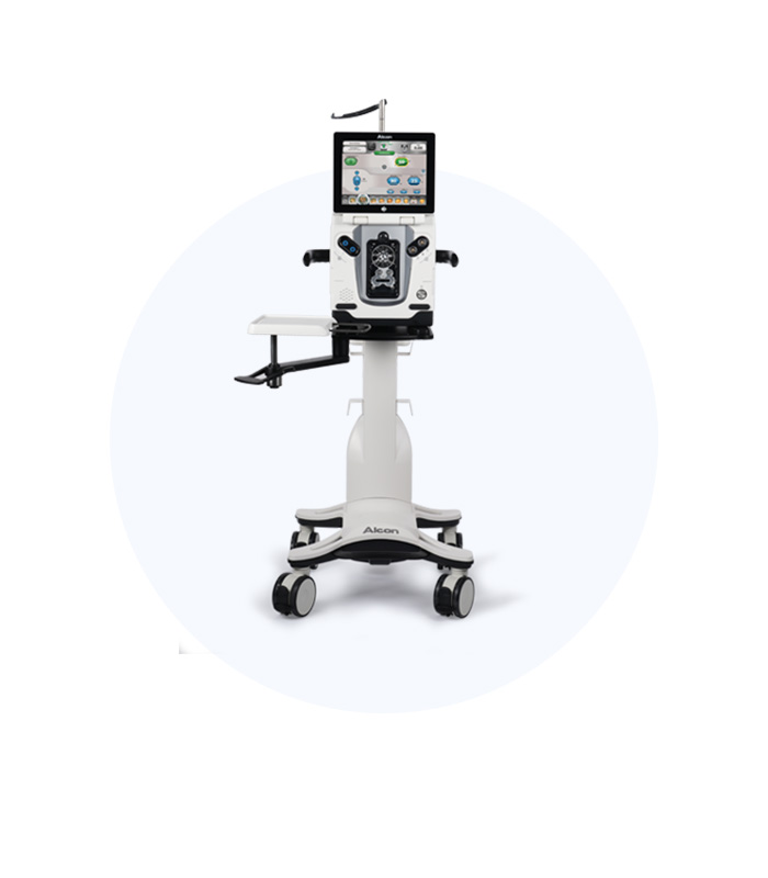
LEGION
LEGION
01. Safe SurgeryThe dual segment technology applied in the FMS of the Legion System reduces fluctuations within the eye, enabling safer surgeries.
02. Rapid Vision RecoveryUsing patented Ozil technology, it reduces heat generation at the incision site, allowing for faster surgeries, which contributes to swift vision recovery.
03. Quick RecoveryWith the capability for 2.2mm small incision surgeries, it allows for rapid recovery of the incision site while minimizing the risk of post-surgical infection.
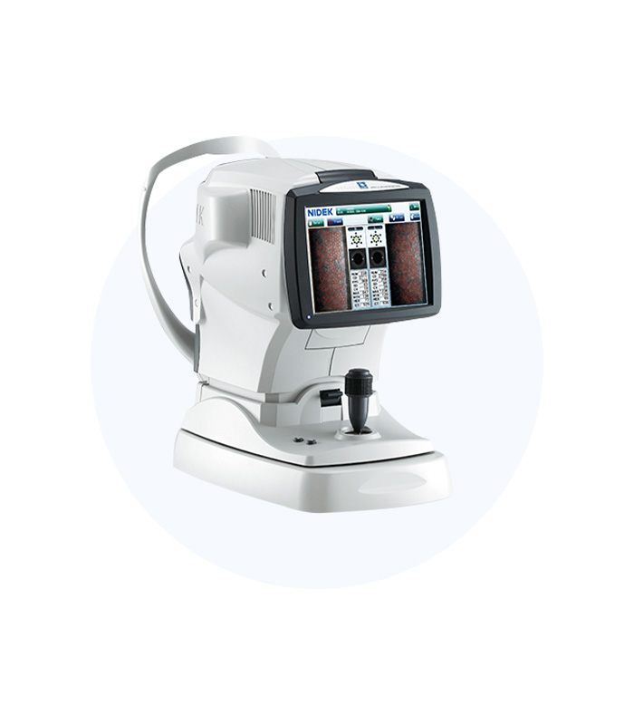
Endothelial Cell Examination Device
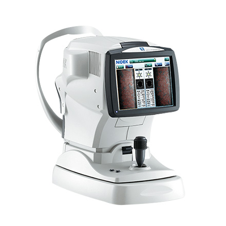
KPREAN SP9000
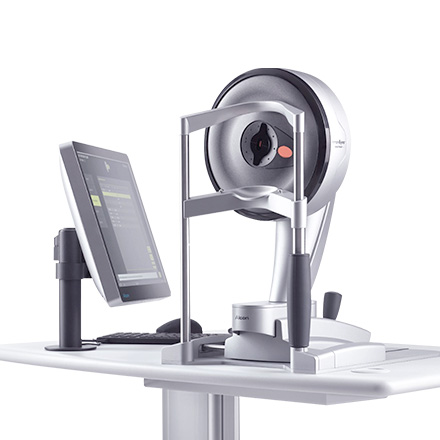
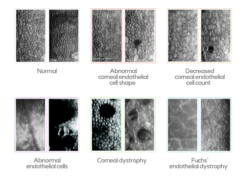
The corneal endothelial cell examination device is a non-contact ophthalmoscope that allows for high-magnification observation of endothelial cells inside the cornea without contact.
It provides critical data for vision correction surgeries, intraocular lens insertions, and cataract surgeries by assessing the number and shape of endothelial cells. The average adult has over 2,000 polygonal endothelial cells, which play a vital role in maintaining corneal transparency.
01. Advantages of Endothelial Cell Examination Devices
The device measures corneal endothelial cells from five directions, allowing for precise examination from the center to the periphery.
Being non-contact, it enables patients to undergo testing comfortably.
It can identify signs of various diseases such as corneal trauma, corneal edema, and corneal dystrophy, providing accurate insights into the corneal condition pre-surgery.
Examination of endothelial cells before and after surgery for candidates of intraocular lens insertion helps select suitable candidates and ensure safe surgical outcomes.
Wide-Angle Camera (Optomap)
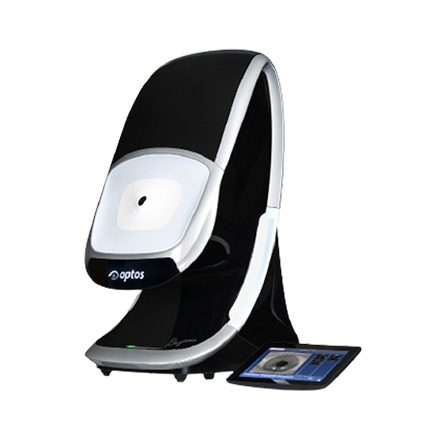
OPTOMAP
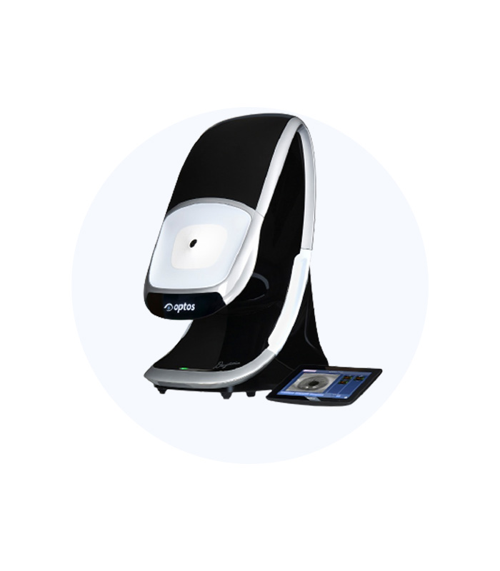
Previous similar devices were limited by pupil size, requiring dilation for examinations that could only capture part of the eye (the macula and optic nerve).
For peripheral examinations, dilation was necessary, and 7 to 15 images had to be stitched together in a panoramic manner, which was inconvenient. However, Optomap allows for wide-area diagnosis in a short time without dilation, enabling the simple detection of retinal and choroidal diseases that could only be confirmed through existing precision tests, facilitating early diagnosis and prevention.
Screens the retina up to 200 degrees (80%) without dilation
Capable of discovering retinal diseases that are difficult to detect with existing devices, allowing for early treatment
Provides a 3D actual image of the patient's retinal condition, increasing trustworthiness
Allows for lifelong management of the patient’s eyes through simple retinal examinations after LASIK surgery.
XTRA
XTRA
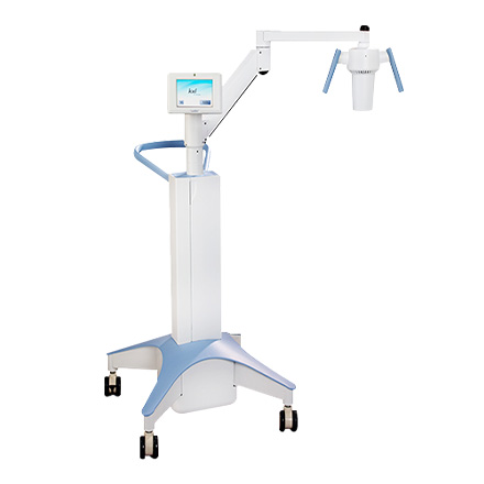
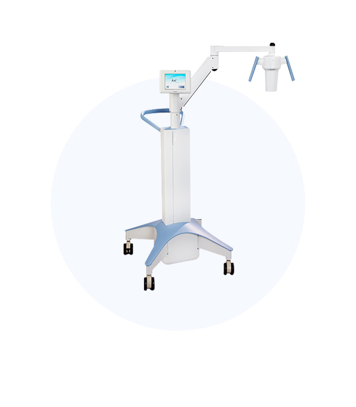
The Avedro KXL system (XTRA) received KFDA approval in October 2014 as a safe collagen cross-linking system.
It strengthens the cornea weakened either congenitally or post-surgery, proving effective in alleviating keratoconus symptoms and is considered safe.
01. Minimized Side Effects through Uniform Energy Delivery
Unlike previous CXL devices that focused energy unevenly on the corneal center, the KXL system provides uniform energy distribution from the center to the periphery, strengthening corneal tissue evenly.
02. CE Mark and KFDA ApprovalLasik/Lasek Xtra® has been consistently performed abroad for several years, with over 80,000 surgeries completed. Avedro KXL has received the CE mark and is in use throughout the EU, as well as approved in Canada, Japan, and Singapore, actively performing surgeries. In this context, the Shinagawa Lasik Center in Japan presented results of over 50,000 Lasik Xtra surgeries at the American Society of Cataract and Refractive Surgery (ASCRS) in 2013, demonstrating long-term stability in corrected vision post-surgery.
03. Features of OCT that Enhance ResultsIn May 2015, at the Avedro Advisory Board held in Zurich, Switzerland, First Samsung Eye Clinic presented on the efficacy and safety of performing collagen cross-linking after LASEK Xtra surgery.
The post-surgical collagen cross-linking provides a lock-in effect on the operated eye, reducing refractive changes and stabilizing vision in the short term compared to regular surgeries. The safe Avedro KXL system approved by KFDA aims for a higher level of safety in vision correction procedures.
PENTACAM
PENTACAM
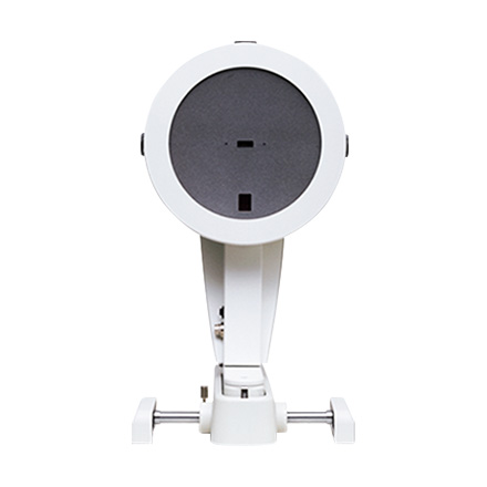
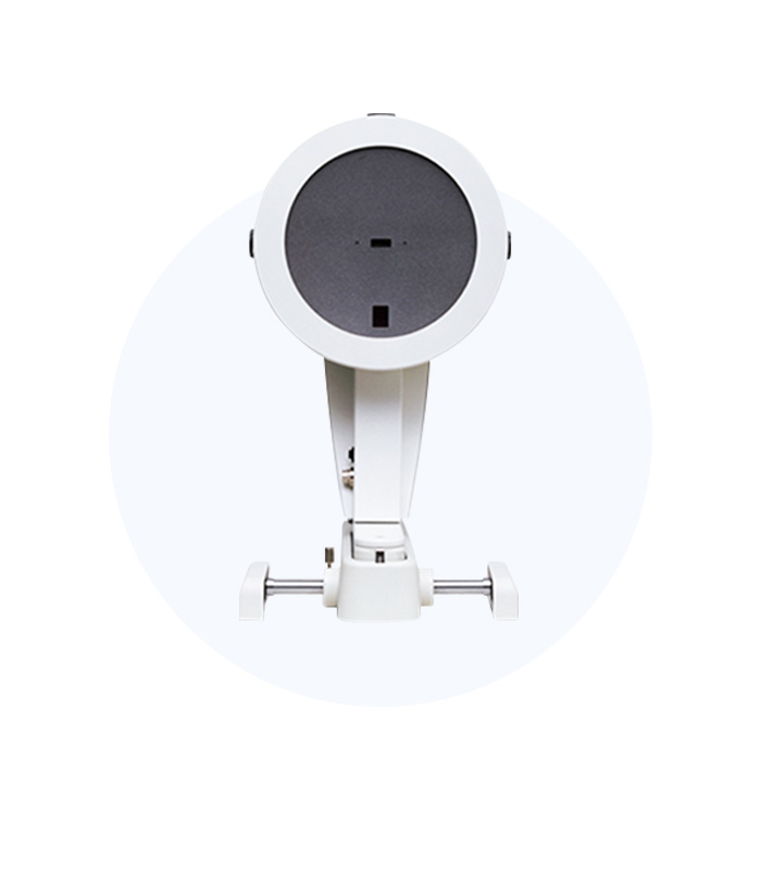
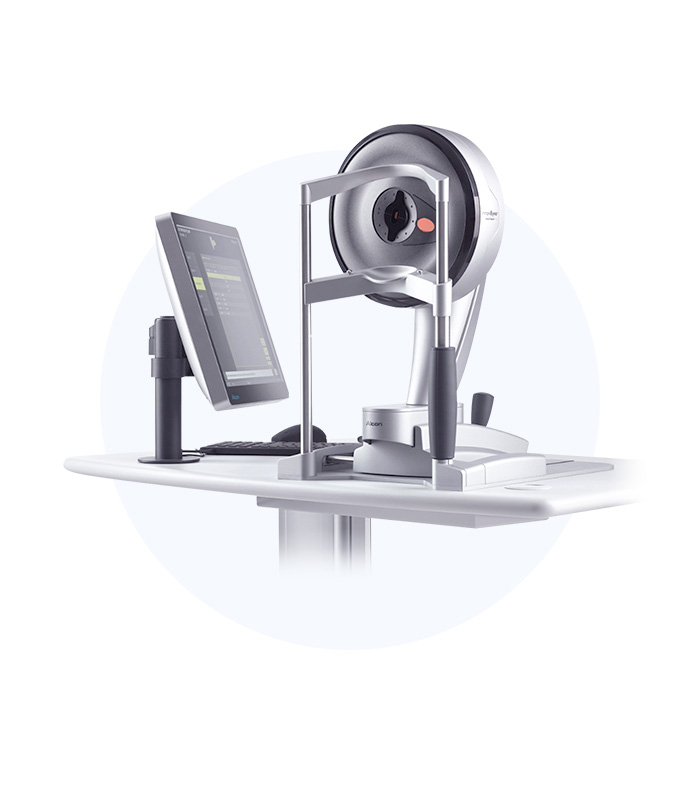
Pentacam is an advanced precision device developed by the German company Oculus, specializing in ophthalmic examination and diagnostic equipment. It uses a Rotating Slit Scanning Camera system to photograph and analyze the anterior segment, including the front and back of the cornea and the lens.
It measures the patient’s fine eye movements simultaneously in 3D, allowing for more precise and accurate examinations than existing anterior segment imaging devices.
It uniquely describes 25,000 elevation points accurately and measures the patient’s subtle eye movements in 3D simultaneously, providing more precise and accurate examinations than existing anterior segment imaging devices.
SITEMAP
SITEMAP
To conduct personalized vision correction surgeries, Personal Eyes examines refraction, irregular astigmatism, and eye length with high precision.
01. Multiple Measurements with One DeviceWavefront measurement of the eye
Measurement and imaging of corneal structure
Biometric measurements (eye size, corneal curvature, and thickness)
Data measured through the sitemap is directly transmitted to the surgical device EX500, eliminating manual input and additional processes, thereby preventing human errors.
Z8
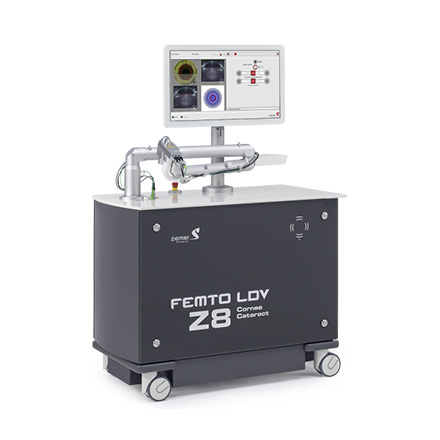
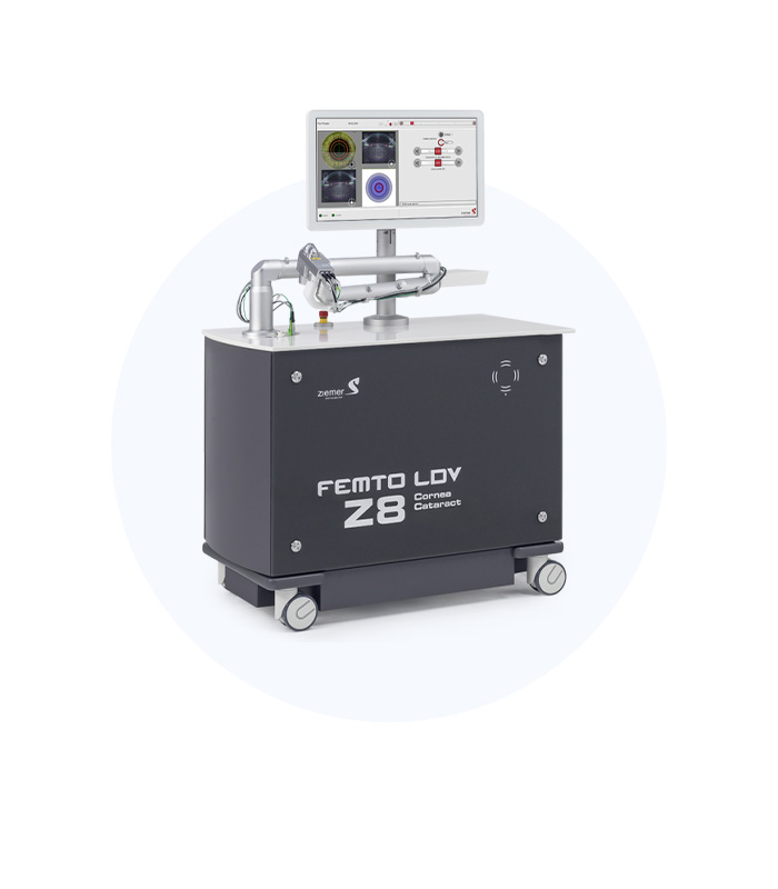
FEMTO LDV Z8
The Z8 is designed, manufactured, and produced in Switzerland. It uses cutting-edge Ziemer nJoule technology to enable ALL-Laser surgery.
01. Safe Surgery through Low Energy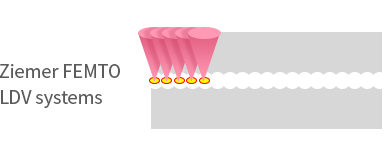
The Cirrus HD-OCT takes images according to the position of the retina based on built-in reference data, ensuring accurate reproduction of previous examinations and providing reliable test results.
02. High Patient Safety
The upgraded Z8 boasts various advantages in safety.
OCT image capture and a high-resolution top view camera
Convenient PI docking
Computer-controlled smart vacuum systems
Additional safety devices (hardstop, interlock, etc.)
Allowing for smooth resections without tissue bridges
Reduced inflammatory responses
No occurrence of pupil dilation due to laser use

깔끔하고 정확한 각막 절개를 통해
백내장 수술 중 매끄러운 난시 교정이 가능하며
절개하는 깊이 및 ARC의 길이를 보다 더 정교하게 설정합니다.
TOPCON DRI OCT Triton
(OCT-A)
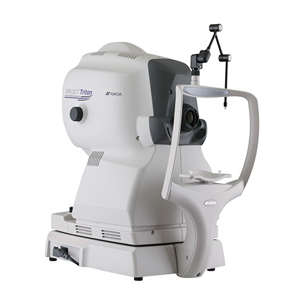
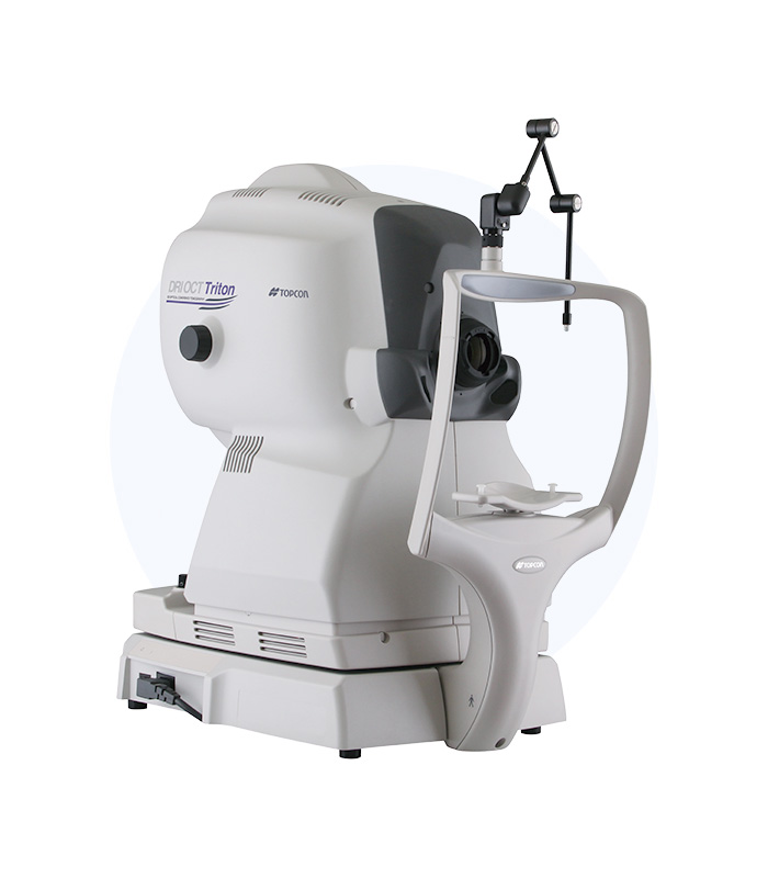
TOPCON DRI OCT Triton(OCT-Angio)
The device functions as an optical coherence tomography (OCT) scanner for the optic nerve and retina, similar to how CT scans are used in comprehensive health checkups. It enables retinal CT scans for comprehensive eye examinations, allowing for the early detection of serious ophthalmologic conditions such as retinal diseases and glaucoma using cutting-edge technology.
OCT imaging, while similar in principle to ultrasound, uses near-infrared light instead of sound waves. By analyzing the quantity and time delay of reflected light from tissues—depending on their properties—using an optical interferometer, it enables high-resolution cross-sectional imaging.
01. ‘Optical Coherence Tomography Angiography’ without Contrast Injection
Equipped with the latest retinal imaging technology, the Optical Coherence Tomography Angiography (OCT-A) system captures the vascular circulation of the retina and choroid, allowing for precise assessment of intraocular vascular conditions.
02. Layer-by-Layer Cross-Sectional ImagingBy capturing vascular images of the retina and underlying choroid in cross-sectional layers, the device enables safer and more convenient observation of internal ocular conditions without missing any details.
03. Key Features of OCT-A for Enhanced ResultsCompared to conventional OCT, OCT-A experiences less interference from cataracts and requires a shorter scanning time, making it possible to conduct more detailed examinations such as retinal and glaucoma screenings simultaneously.
Topcon's High-Resolution Optical Technology
- High-resolution, layer-specific imaging produces clear visual data, enhancing diagnostic reliability.
Database for All Ethnicities
- Equipped with a Normative Database inclusive of both Eastern and Western populations, enhancing examination convenience and intuitive usability.
3D Panoramic Positional Analysis Function
- Automatically analyzes and adjusts vascular positioning during scanning, increasing the reliability of longitudinal disease tracking data.
