Conical cornea
-
Eye Disease Clinic
-
Conical cornea
What is a
conical cornea?
It is a progressive disease in that parts of the cornea
are getting thinner and thinner, causing them to
protrude forward instead of maintaining their
original gently rounded shape.
The cause is not yet clearly identified, but rubbing
the eye is thought to be the most important factor.
More recently, it has been reported as a side effect
of LASIK surgery, and conical cornea,
which is also known as keratoconus.
When too much of the residual cornea is removed
during LASIK surgery without leaving enough for safety,
the cornea is unable to maintain its original shape
and protrudes forward.

Conical Cornea Symptoms
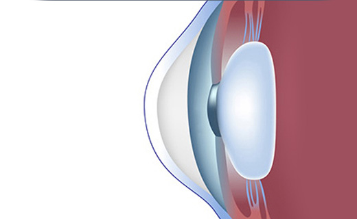
Normal Cornea
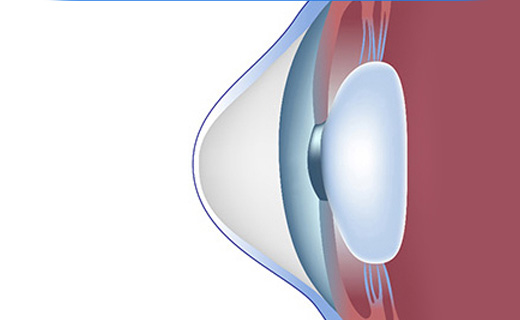
Conical cornea
In the early stages of conical cornea, there are no significant abnormalities in vision or only subtle changes are noticed.
As the disease progresses, the cornea becomes thinner and more protruding,
distorting the light that enters the eye and causing vision loss due to irregular astigmatism, as well as glare and light flashes.
If your vision is deteriorating and changing your glasses or contact lenses within a short period of time,
you may have conical cornea, and an eye clinic visit is recommended.
Conical cornea ring implant treatment
at First Samsung Eye Clinic
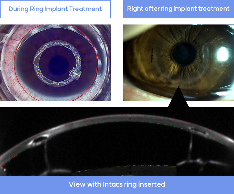
Ring implant for conical cornea
The laser makes a safe tunnel for
the ring in 6 seconds.
The treatment takes only about five minutes.
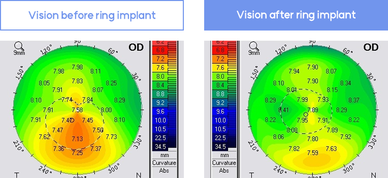
First Samsung Eye Clinic Cases
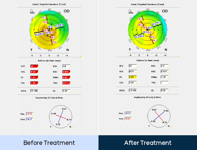
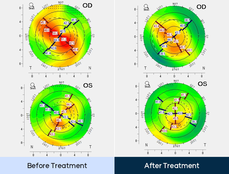
These are before(Left, Vision 0.6) and
after(Right, Vision 1.0) of ring implantation.
If conical cornea has found in early like this patient,
you can keep your vision as normal with ring implantation.
Corneal Crosslinking Treatment
at First Samsung Eye Clinic
Corneal Crosslinking Surgery
Corneal Crosslinking is an essential treatment for all
conical corneas because it makes the cornea four times stronger.
A single treatment can prevent most conical cornea from
progressing, avoiding keratoplasty.
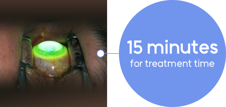
First Samsung Eye Clinic offers the latest treatment,
which reduces the treatment time from one hour to 15 minutes.
Simultaneous treatment of
early myopia and astigmatism
Corneal Crosslinking + LASEK
Performing Corneal Crosslinking and LASEK allows
early myopia and astigmatism to be treated at the same
time as the conical cornea.
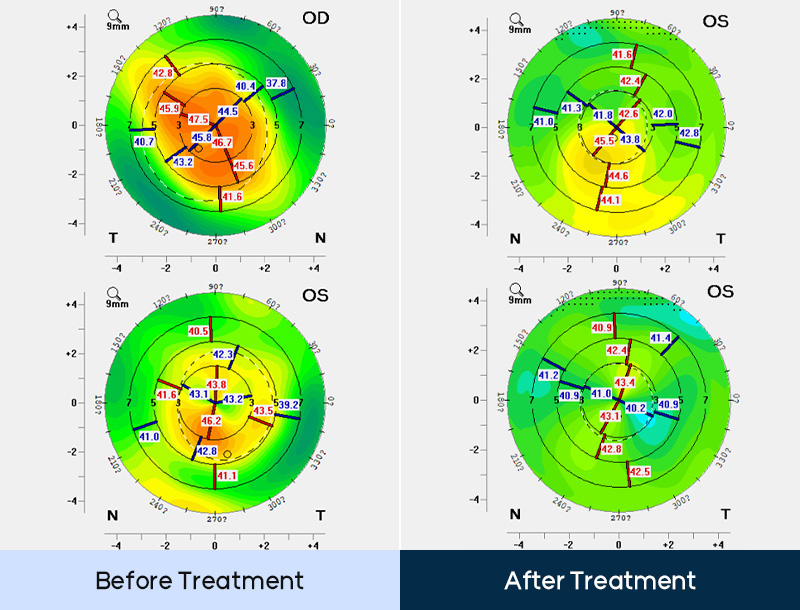
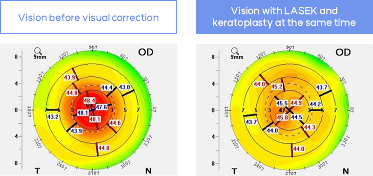
The centrally located conical cornea (left)
resulted in a corrected vision of only 0.2.
By performing a special LASEK
(corneal topography-based LASEK) and keratoplasty
at the same time, it improved to 0.8 at 3 months
postoperatively (right).
Conical Cornea Treatment Cases
at First Samsung Eye Clinic
First Samsung Eye Clinic YOUTUBE


